Every MRI fistulogram report, whatever the radiologist’s style, answers the same six questions: where the tract starts (the internal opening), where it runs, how it relates to the sphincter muscles, whether there are secondary tracts, whether there is an abscess, and what grade the radiologist assigns. Once you can find those six lines, you can understand what the scan changes in your operation.
Almost every fistula patient at Sharvari Hospital is imaged before surgery — an MRI fistulogram or a trans-rectal ultrasound. The scan is not a formality. The operation follows the tract, and the report is where the tract is written down. This guide walks through the six lines in the order they appear on most reports, then reads a real, de-identified report with you.
The six lines every MRI fistulogram report has
1. Internal opening
This is the point inside the anal canal where the tract begins. It is reported as a clock position — 12 o’clock is the front, 6 o’clock is the back, 3 o’clock is your left — and usually a level, such as “lower anal canal” or “at the dentate line”. Every fistula operation has to reach and deal with this opening. A missed internal opening is the commonest reason a fistula comes back after surgery.
2. The tract
The report describes the tunnel itself: where it goes, how long it is, how wide it is, whether it is straight or curved, and which fat space it passes through (the ischioanal fossa is the fat on either side of the anus). You may also see a description of the tract’s signal — “hyperintense on STIR” simply means the tract lights up on the sequence that shows fluid and inflammation, which is how the radiologist sees it at all.
3. Relation to the sphincters
This is the single most important line. Two rings of muscle surround the anal canal: the internal sphincter, which works on its own, and the external sphincter, which is the muscle you squeeze to hold stool. The report will say the tract is intersphincteric (runs between the rings and never crosses the outer one), trans-sphincteric (passes through both rings), or, less commonly, suprasphincteric or extrasphincteric (climbs above the pelvic floor or bypasses the sphincter entirely). The word decides whether the tract can be laid open or must be closed in a muscle-sparing way.
4. Secondary tracts and horseshoe extension
A secondary tract is a branch off the main tunnel. A horseshoe extension is a tract that curves around the back of the anal canal to the other side. Both are easy to miss with a probe alone and are a common reason a first operation “did not work”. The report will say “no secondary tract” or will describe where the branch runs.
5. Abscess or collection
If there is a pocket of pus along the tract, the report will call it an abscess or a collection and give its size. This changes the order of surgery: infection is drained first, and the definitive procedure follows once the tissue has settled.
6. Grade
Many reports end with a grade from the St James’s University Hospital MRI classification — a shorthand from 1 to 5 that summarises the lines above. Grades 1 and 2 are intersphincteric (2 with an abscess or branch); grades 3 and 4 are trans-sphincteric (4 with an abscess or branch); grade 5 extends above the levator muscle. The full table is on our MRI fistula mapping page.
What each line means for you
| Line on the report | What it changes |
|---|---|
| Internal opening | The point the operation must reach; a clear position makes the probe’s job predictable |
| Tract length and course | How much tissue is involved and which route the surgeon takes |
| Intersphincteric | Usually safe to lay open (fistulotomy) |
| Trans-sphincteric | The part inside the external sphincter is preserved — FiLaC laser, LIFT or a seton for that segment; a hybrid of lay-open plus laser is common |
| Supra- or extrasphincteric | Never laid open; seton, flap or a staged plan |
| Secondary tract or horseshoe | Every branch must be addressed; often a staged plan |
| Abscess | Drained first; definitive surgery later |
| Grade | A summary for the radiologist’s records — the plan is built from the findings, not the number |
A real case, line by line
The report below belongs to a man in his twenties who came to Sharvari Hospital with perianal pain and discharge. It is reproduced as findings only, with every identifying detail removed, and shared with his written consent.
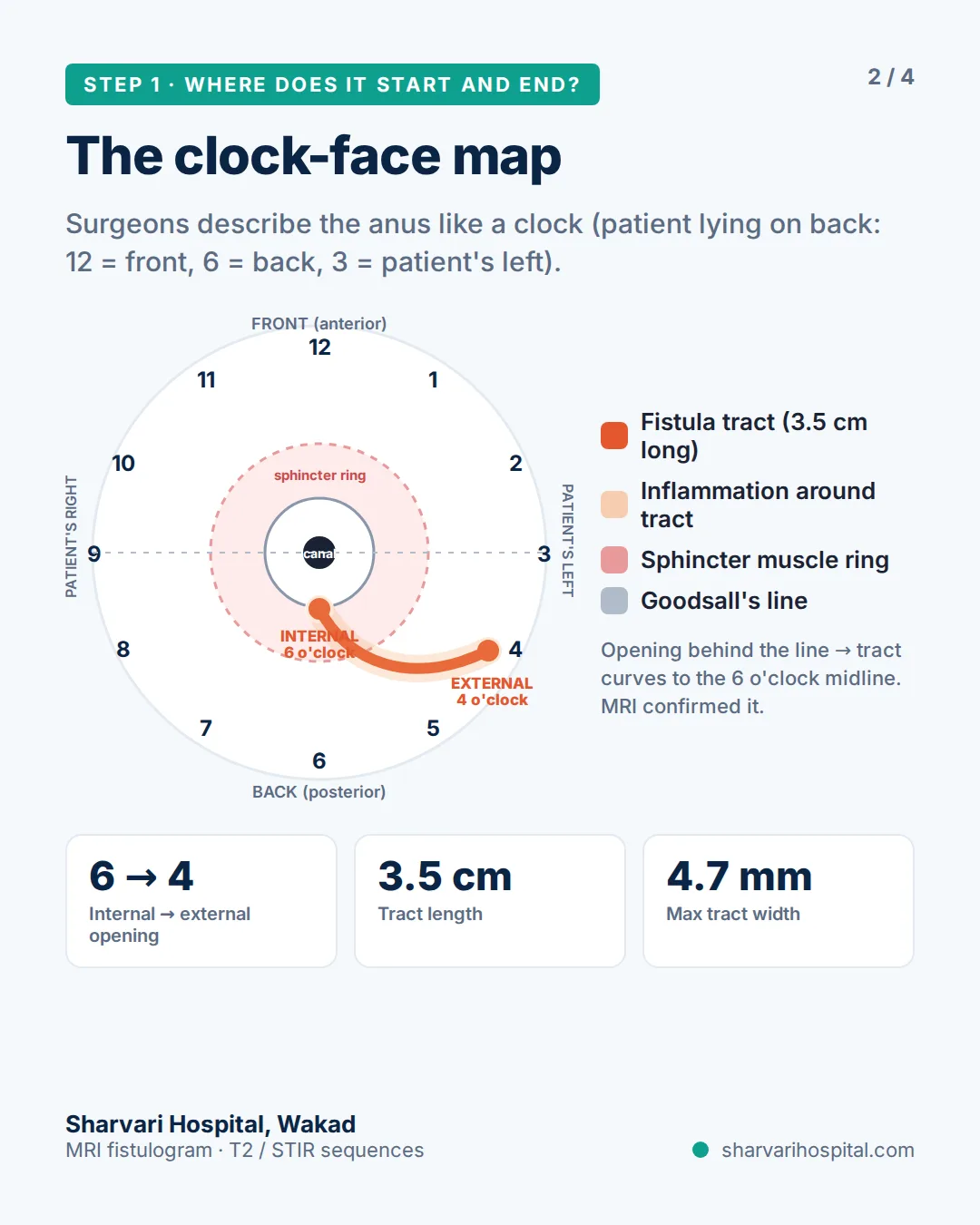
Line 1 — internal opening: 6 o’clock, lower anal canal. The tract starts in the posterior midline, exactly where Goodsall’s rule predicts for an external opening behind the anus.
Line 2 — tract: a single tract, 3.5 cm long and 4.7 mm at its widest, curving through the left ischioanal fossa from the internal opening to an external opening at 4 o’clock, with mild inflammation along its length.
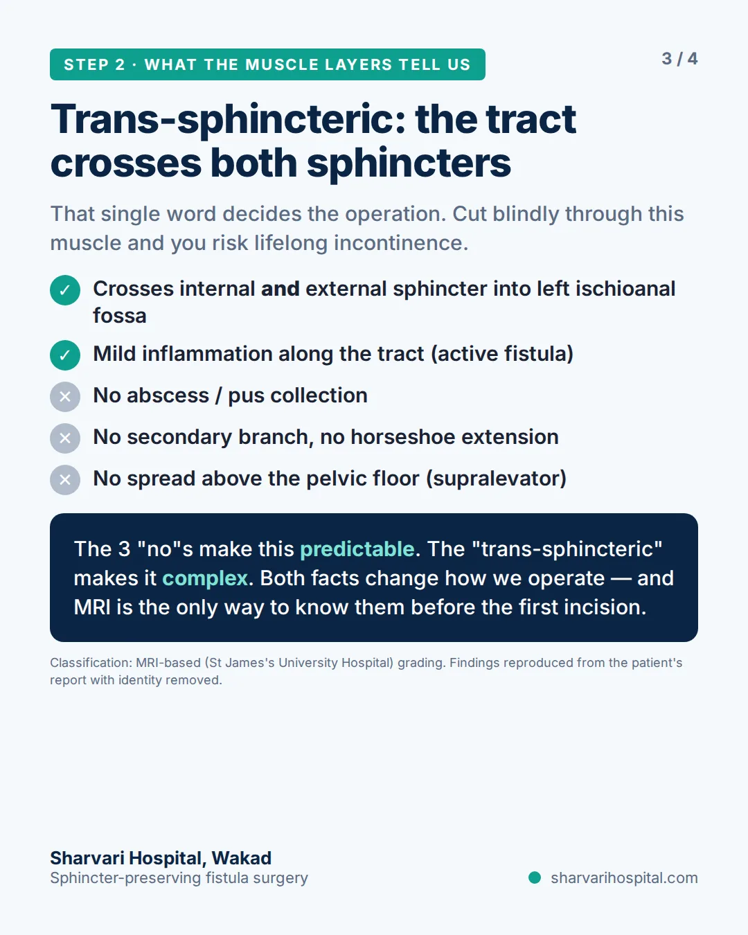
Line 3 — sphincters: trans-sphincteric. The tract crosses both the internal and the external sphincter on its way out. That one word is what turned a “simple-looking” fistula into a complex one.
Lines 4 and 5 — branches and abscess: none. No secondary tract, no horseshoe extension, no collection, no spread above the pelvic floor. Three “no” findings made this a predictable single tract.
Line 6 — grade: the report carried a grade, but the operation was planned from lines 1 to 5, not from the number. That is the honest position on every report: the grade is a summary, the findings are the plan.
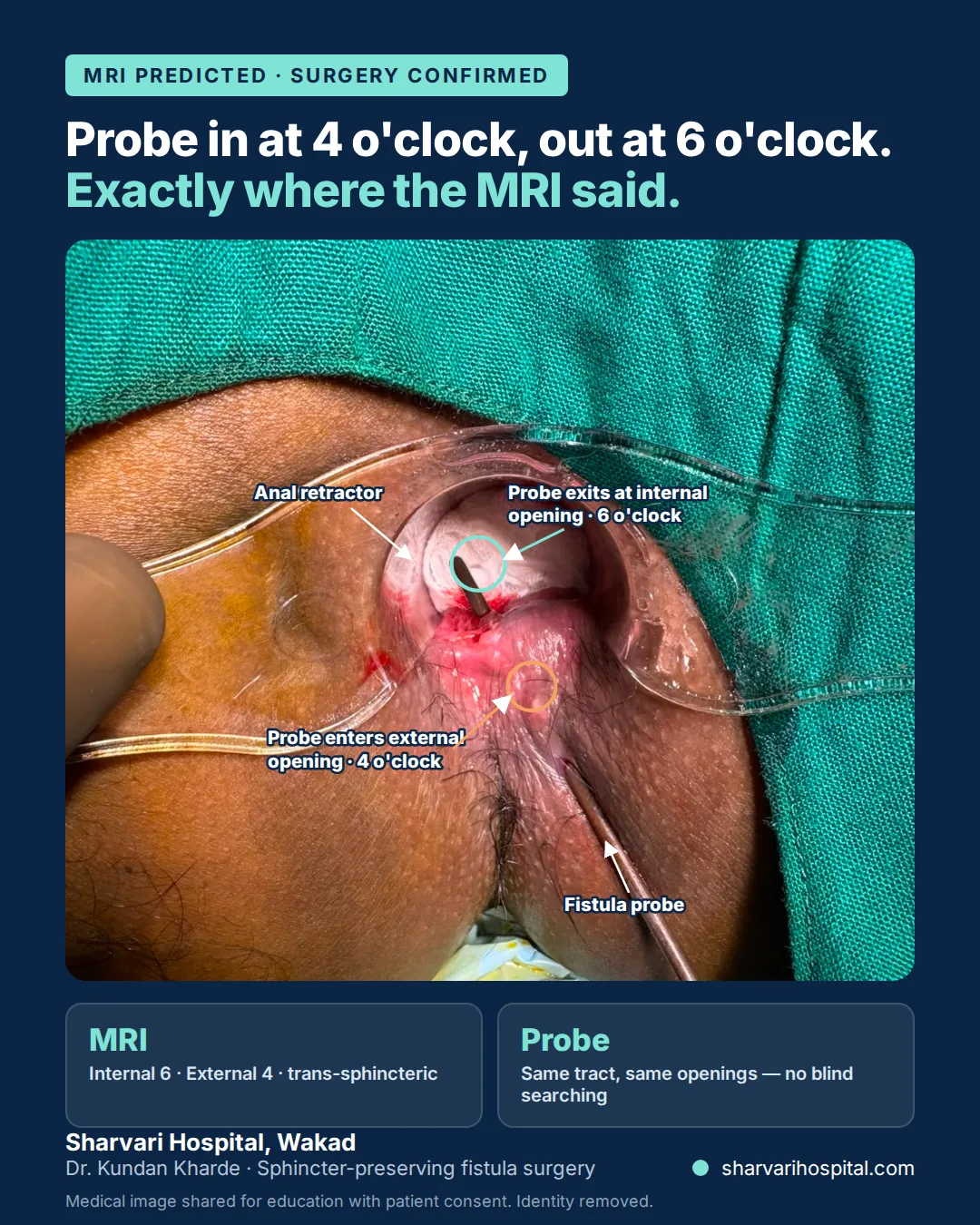
In theatre the probe went in at 4 o’clock and came out at 6 o’clock along the same tract — the scan and the probe agreed, so nothing was searched for blindly.
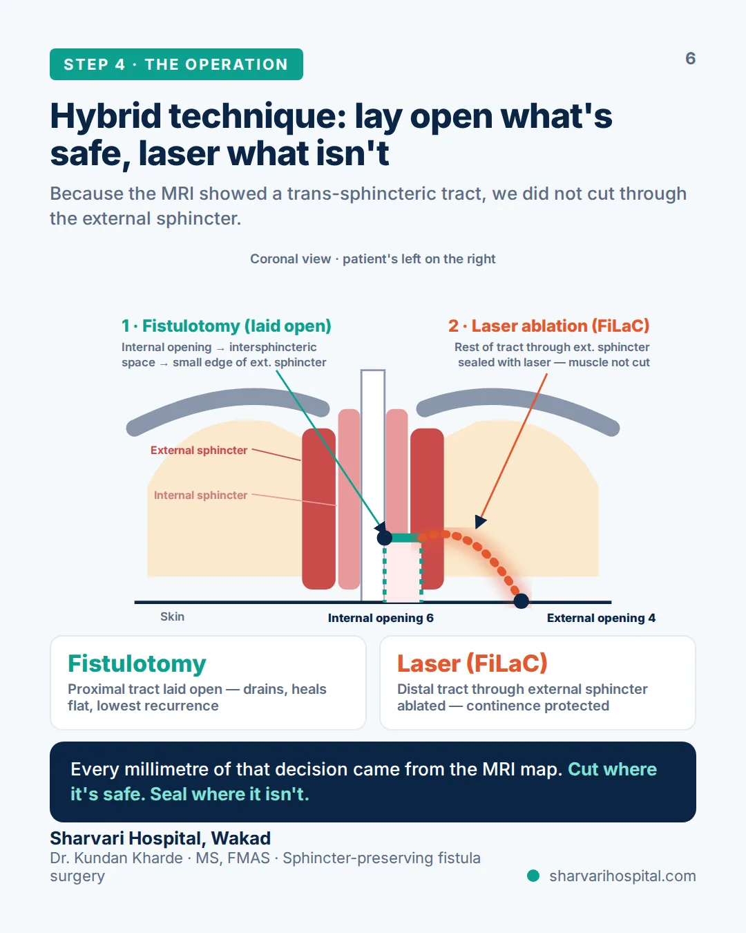
Because line 3 said trans-sphincteric, the whole tract was not laid open. The part from the internal opening through the intersphincteric space was opened, taking only a small edge of the external sphincter; the remainder, running through the external sphincter, was sealed from inside with FiLaC laser so the muscle was not divided. The complete write-up is on the case study page.
Five questions to ask your surgeon after the MRI
- Where exactly is my internal opening, and how will you confirm it in theatre? The answer should be a clock position and a method (probe, dye or both).
- How much of my external sphincter does the tract cross? This decides whether any part of the tract can be laid open.
- Did the scan show any secondary tract, horseshoe extension or abscess? If yes, ask how each one will be dealt with — and whether treatment will be staged.
- Which operation are you planning from these findings, and what is the alternative? A surgeon reading the map should be able to explain why a fistulotomy, a hybrid, LIFT, FiLaC or a seton fits this particular tract.
- What will change if the findings in theatre differ from the scan? The MRI is a map, not a contract; the plan is confirmed under anaesthesia.
What to do next
If you already have an MRI fistulogram, you do not need to interpret it alone. Send the written report and photos of the key images to +91 951 951 1928 on WhatsApp and Dr. Kundan Kharde will read it before you travel. For the full explanation of the clock-face map, the muscle layers and the St James’s grades, read MRI fistula mapping before surgery. First-time patients can start at fistula treatment in Pune; if your fistula has come back after an operation, the complex and recurrent fistula page describes how a redo is planned.
Frequently asked questions
What does ‘internal opening at 6 o’clock’ mean on my MRI report?
Positions around the anus are described like a clock face with the patient lying on the back: 12 o’clock is the front, 6 o’clock is the back towards the tailbone, and 3 o’clock is the patient’s left. An internal opening at 6 o’clock means the tract starts in the posterior midline of the anal canal, which is the commonest site.
What does ‘trans-sphincteric’ mean on an MRI fistulogram?
It means the tract passes through both the internal and the external anal sphincter on its way to the skin. Part of the tract sits inside the muscle that controls stool, so that part cannot simply be cut open; the surgeon plans a sphincter-preserving step such as FiLaC laser, LIFT or a seton for it.
Is a higher St James’s grade worse?
A higher grade means the tract runs further from the anal canal, or has an abscess or a secondary branch, so the operation is usually more involved. The grade is a radiology shorthand; the operation is planned from the actual findings on the scan and confirmed under anaesthesia, not from the number alone.
Can I get an opinion on my MRI fistulogram before visiting Sharvari Hospital?
Yes. WhatsApp the written report and photos of the key images to +91 951 951 1928 and Dr. Kundan Kharde will review them before you travel. A clinical examination is still needed before any operation is confirmed, so the WhatsApp opinion is a starting point rather than a final plan.
Related Reading
- MRI Fistula Mapping in Pune — Every Complex Fistula Mapped Before Surgery
- Case study: trans-sphincteric fistula mapped on MRI, treated with hybrid fistulotomy + FiLaC
- How Is Anal Fistula Diagnosed? MRI, Ultrasound and Tests Explained
- Types of Anal Fistula: Simple vs Complex
- Questions to Ask Before Fistula Surgery
- Complex & Recurrent Fistula Treatment →
Written and reviewed by Dr. Kundan Kharde, MBBS, MS – General Surgery, FMAS, Sharvari Hospital, Pune. Last reviewed: September 2026.
This article is for patient education only and does not replace an in-person medical consultation. The case described is de-identified and shared with the patient’s written consent; no outcome is promised from a single case.