Imaging-first fistula surgery · Pune
MRI Fistula Mapping in Pune — Every Complex Fistula Mapped Before Surgery
An MRI fistulogram is a pelvic MRI scan done with a fistula protocol. It shows the whole tunnel of an anal fistula — where it starts inside the anal canal, the path it takes, how much sphincter muscle it crosses, and whether there are hidden branches or a pocket of pus — before anyone operates. Almost every fistula patient at Sharvari Hospital is imaged before surgery — an MRI fistulogram or a trans-rectal ultrasound.
The reason is simple: the operation follows the tract. A missed internal opening is the commonest reason a fistula comes back after surgery, and the amount of sphincter the tract crosses decides whether muscle can be cut at all. The map is made first; the operation is chosen from it.
Book Consultation
Get a callback within 30 minutes
What an MRI fistulogram shows
Five findings on the scan decide the operation. A clinical examination can suggest some of them; only imaging shows all five together.
- 1
The internal opening — where the tract starts inside the anal canal — the point every operation must reach. Missing it is the commonest reason a fistula comes back.
- 2
The course and length of the tract — whether it runs straight or curves, how far it travels, and how wide it is.
- 3
Sphincter involvement — how much of the internal and external sphincter muscle the tract crosses — this decides whether muscle can be cut or must be preserved.
- 4
Secondary tracts and horseshoe extensions — side branches or a tract that loops around the anal canal, which a probe alone can miss.
- 5
Abscess and supralevator extension — a pus collection that must be drained first, or spread above the pelvic floor that changes the whole approach.
The clock-face map, explained
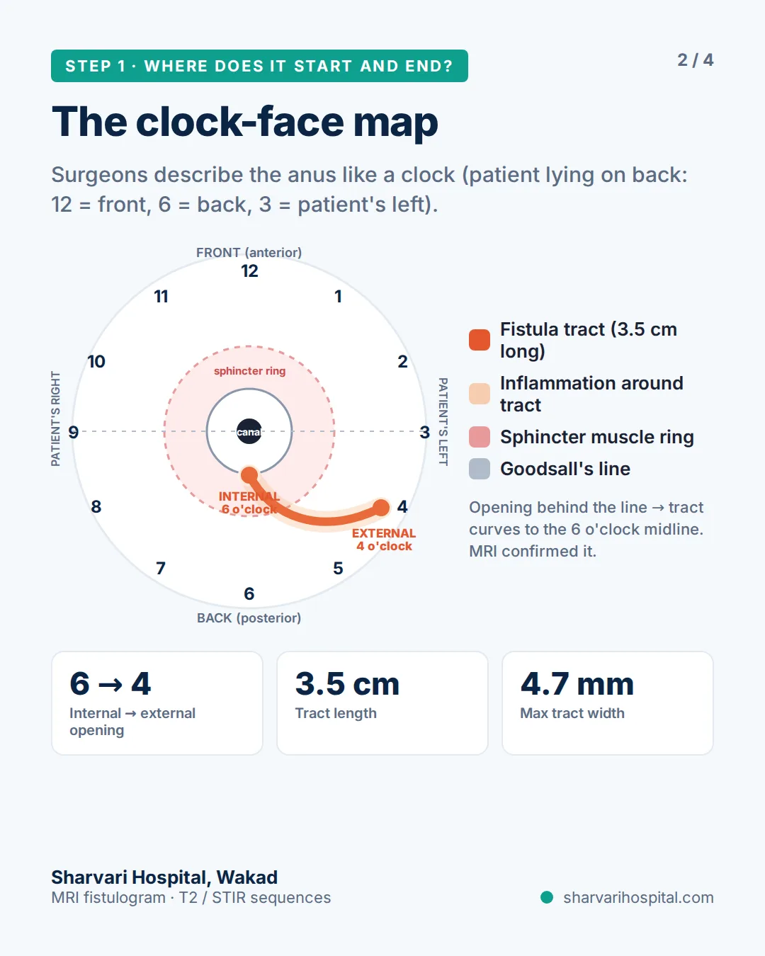
Surgeons and radiologists describe positions around the anus like a clock face, with the patient lying on the back in the lithotomy position: 12 o’clock is the front (towards the scrotum or vagina), 6 o’clock is the back (towards the tailbone), and 3 o’clock is the patient’s left. When your report says “internal opening at 6 o’clock”, it means the tract starts in the posterior midline of the anal canal.
Goodsall’s rule is the old surgical shortcut for predicting the route: an external opening in front of an imaginary line drawn across the anus usually has a straight tract to the nearest point of the canal, while an external opening behind that line usually curves round to an internal opening in the posterior midline at 6 o’clock. The rule is a useful guess, not a map — the case on this page had an external opening at 4 o’clock, behind the line, and the MRI confirmed the curve to 6 o’clock before surgery rather than assuming it.
How the muscle layers decide the operation
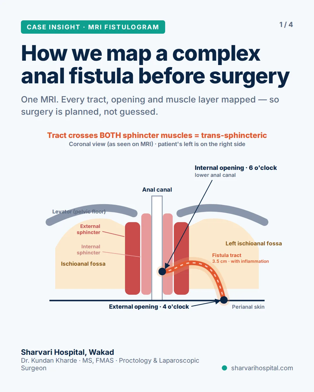
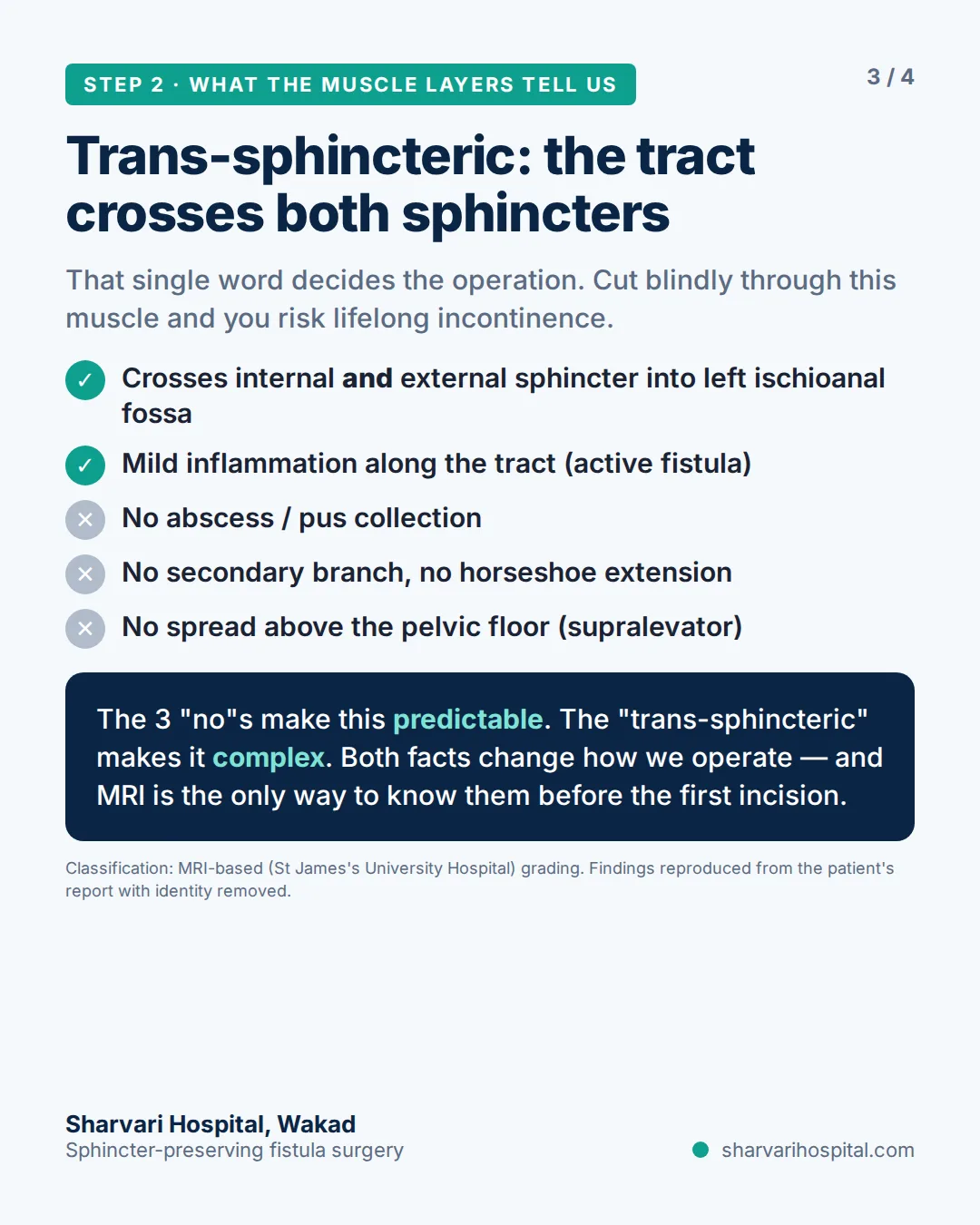
Two rings of muscle surround the anal canal. The inner ring (internal sphincter) works on its own; the outer ring (external sphincter) is the muscle you squeeze to hold stool. Every fistula tract is described by how it relates to these rings:
- Intersphincteric — the tract runs between the two rings and reaches the skin without crossing the outer muscle. Most of these can be opened safely.
- Trans-sphincteric — the tract passes through both rings on its way out. Part of it sits inside the muscle you need for control, so that part cannot simply be cut.
- Supra- or extrasphincteric — the tract climbs above the pelvic floor or bypasses the sphincter entirely. These are never laid open.
| MRI finding | What it changes in surgery | Why |
|---|---|---|
| Intersphincteric tract, low, no branch | Fistulotomy (lay-open) | The tract crosses little or no external sphincter, so it can be opened along its length and left to heal flat. |
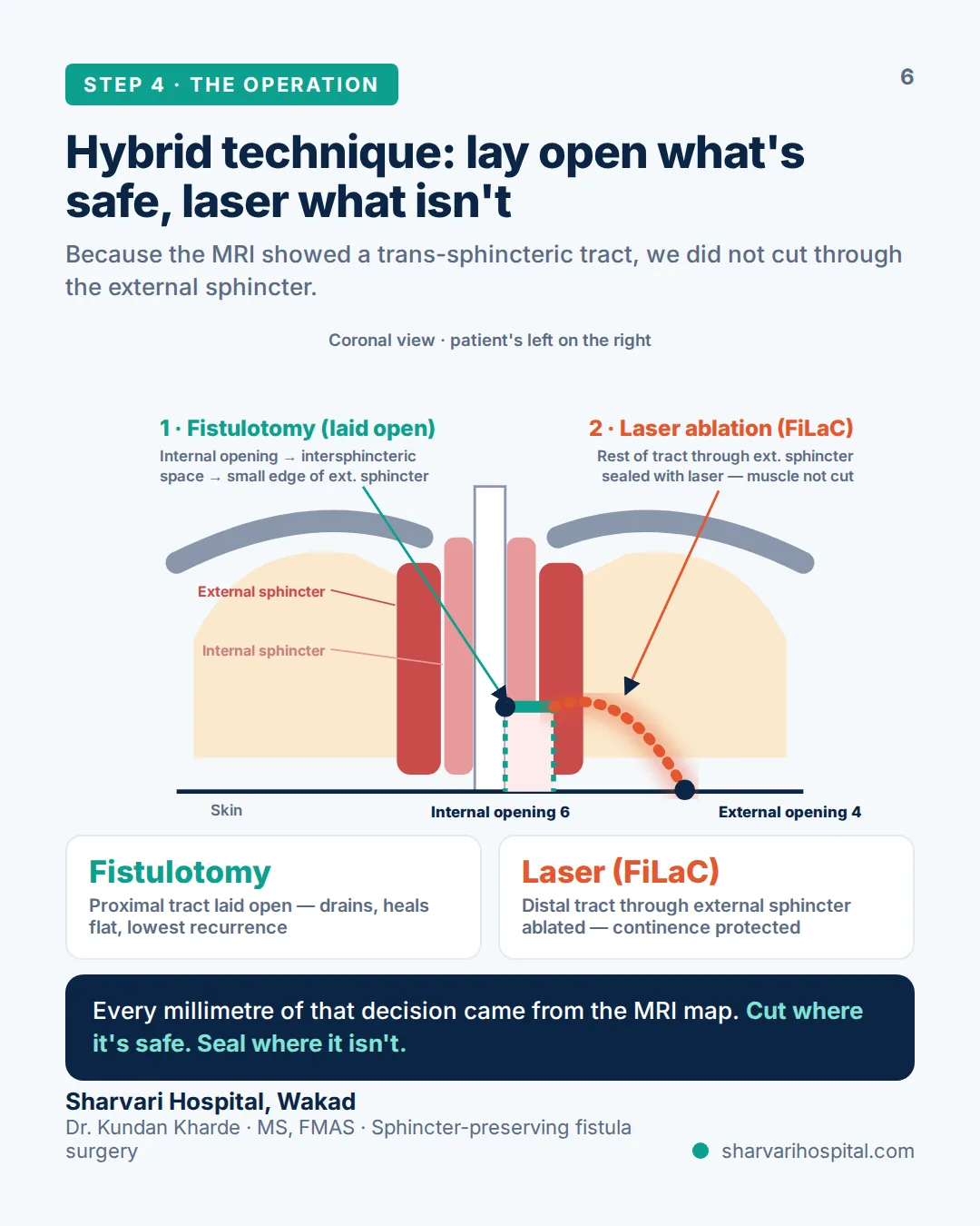
| Trans-sphincteric tract crossing part of the external sphincter | Hybrid fistulotomy + FiLaC | The part of the tract that is safe to open is laid open; the part inside the external sphincter is sealed with laser so the muscle is not divided. |
| High trans-sphincteric tract (a large share of the external sphincter) | LIFT or FiLaC alone, often after a draining seton | Too much muscle is involved to cut; the tract is closed between the muscle planes or ablated from inside. |
| Abscess or active inflammation around the tract | Drainage or a draining seton first; definitive procedure later | Infected tissue does not hold a repair. The seton controls sepsis and lets the tract settle. |
| Secondary tract or horseshoe extension | Staged plan that addresses every branch | Treating the main tract while a branch is left behind is a recurrence waiting to happen. |
| Supra- or extrasphincteric extension | Never a fistulotomy — seton, advancement flap or staged sphincter-preserving repair | The tract runs above or outside the sphincter complex; laying it open would divide the whole muscle. |
First-time fistula → Fistula treatment in Pune · Recurrent or already-operated fistula → Complex & recurrent fistula treatment
Reading the grade on your report (St James’s University Hospital MRI classification)
Many MRI reports end with a grade. It is a radiology classification that summarises where the tract runs and whether an abscess or branch is present.
| Grade | What it means in plain English |
|---|---|
| Grade 0 | Normal appearance — no fistula tract seen. |
| Grade 1 | Simple, straight intersphincteric tract between the two sphincter muscles, with no branch and no abscess. |
| Grade 2 | Intersphincteric tract with an abscess or a secondary branch, still confined between the sphincters. |
| Grade 3 | Trans-sphincteric tract — crosses both sphincters into the ischioanal fossa, with no abscess and no branch. |
| Grade 4 | Trans-sphincteric tract with an abscess or a secondary tract in the ischioanal fossa. |
| Grade 5 | Tract extending above the levator muscle (supralevator) or passing through it (translevator). |
Grades are a radiology shorthand; the surgical plan is made from the actual findings and confirmed in the operating theatre.
A real case: mapped on MRI, confirmed with the probe
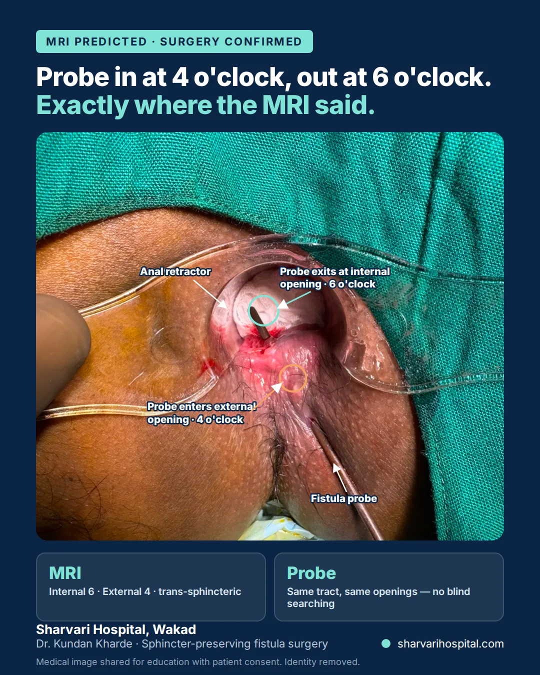
A man in his twenties came to Sharvari Hospital with perianal pain and discharge. Examination found an external opening on the left, behind the anus. His MRI fistulogram showed a single tract starting at the internal opening at 6 o’clock and curving through the left ischioanal fossa to the external opening at 4 o’clock — 3.5 cm long and 4.7 mm at its widest. The tract crossed both the internal and the external sphincter, making it trans-sphincteric, with mild inflammation along its length. There was no abscess, no secondary tract, no horseshoe extension and no spread above the pelvic floor. In theatre the probe entered at 4 o’clock and came out at 6 o’clock along the same tract — exactly as the scan had predicted. Because the scan had shown muscle involvement, the plan was a hybrid: the part of the tract that was safe to open was laid open, and the part running through the external sphincter was sealed with FiLaC laser so the muscle was not divided.
MRI fistulogram or trans-rectal ultrasound — which one?
Almost every fistula patient at Sharvari Hospital is imaged before surgery — an MRI fistulogram or a trans-rectal ultrasound. Which of the two is chosen depends on what the examination suggests.
Trans-rectal ultrasound (TRUS) is usually enough for:
- ✓Short, low tracts where the external opening is close to the anal verge
- ✓A first-time, uncomplicated fistula with no history of abscess surgery
- ✓When MRI is not possible — a pacemaker or other implant, severe claustrophobia, or when the scan cannot be arranged
MRI fistulogram is preferred for:
- ✓A fistula that has come back after previous surgery
- ✓A high tract, or one suspected to involve a large share of the external sphincter
- ✓Suspected branching, horseshoe extension or a collection on examination
- ✓Any previous anorectal surgery that has changed the anatomy
- ✓Suspected Crohn’s disease or tuberculosis as the underlying cause
How is anal fistula diagnosed? MRI, ultrasound and tests explained
MRI fistulogram cost in Pune
A pelvic MRI with fistula protocol at imaging centres in Pune generally costs in the region of ₹4,000 to ₹10,000. This is an indicative market range at partner imaging centres, not a Sharvari Hospital price; the figure varies with the centre, the magnet strength, the sequences run and whether contrast is used. The scan is done at an imaging centre and the report and images come to the hospital for planning.
If you have health insurance, ask your insurer or TPA whether pre-operative investigations are covered under your policy — many policies include them when surgery follows within a set period. Surgery itself is cashless with 50+ insurers at Sharvari Hospital.
Fistula surgery cost in Pune 2026 · Fistula treatment insurance & cashless in Pune
Already have an MRI? Get Dr. Kharde’s opinion on it
WhatsApp the written report and photos of the key images to +91 951 951 1928. Dr. Kundan Kharde reads it before you travel and tells you what the findings mean for the operation.
Fistula that has come back after surgery is planned on its own page: complex & recurrent fistula treatment. First-time patients start at fistula treatment in Pune.
Your surgeon: Dr. Kundan Kharde, MS, FMAS — Founder & Director, Lead Proctology Surgeon, Sharvari Hospital. 19+ years, 6,000+ surgeries.
Frequently asked questions
Do I need an MRI before fistula surgery?
Almost every fistula patient at Sharvari Hospital is imaged before surgery — an MRI fistulogram or a trans-rectal ultrasound. In the case shown on this page, the MRI mapped a trans-sphincteric tract from the 6 o’clock internal opening to a 4 o’clock external opening, and the probe in theatre confirmed it exactly — which is why the plan was a hybrid fistulotomy with FiLaC rather than a full lay-open.
What does "trans-sphincteric" mean on my MRI report?
It means the fistula tract passes through both the internal and the external anal sphincter — the two muscle rings that control stool — before reaching the skin. That single word decides the operation: a trans-sphincteric tract cannot simply be laid open along its whole length without dividing muscle, so the surgeon plans a sphincter-preserving step such as FiLaC, LIFT or a seton for the part inside the external sphincter.
What is the St James’s grade on my MRI report?
The St James’s University Hospital classification is a radiology shorthand that grades an anal fistula from 1 to 5 by where the tract runs relative to the sphincters and whether there is an abscess or a secondary tract. Grades 1 and 2 are intersphincteric; grades 3 and 4 are trans-sphincteric; grade 5 extends above the levator muscle. The surgical plan is made from the actual findings, not from the number.
Can an MRI show why my fistula came back after surgery?
Often, yes. A recurrent fistula usually means an internal opening that was never found, a secondary tract or horseshoe extension that was left behind, or an undrained collection. MRI shows all three, along with scarring from the previous operation and how much sphincter muscle is still intact — which is why a fresh MRI fistulogram is the starting point for any redo fistula surgery at Sharvari Hospital.
How much does an MRI fistulogram cost in Pune?
A pelvic MRI with fistula protocol at imaging centres in Pune generally costs in the region of ₹4,000 to ₹10,000, depending on the centre, the magnet strength and whether contrast is used. This is an indicative market range, not a Sharvari Hospital price. If you have health insurance, ask your insurer whether pre-operative investigations are covered under your policy — many policies include them when surgery follows.
Can I send my MRI report on WhatsApp for an opinion before visiting?
Yes. Send the written report and, if possible, photos of the key images or the film to +91 951 951 1928 on WhatsApp. Dr. Kundan Kharde reviews the report before you travel and tells you whether the findings point to a simple lay-open, a sphincter-preserving procedure or a staged plan. A clinical examination is still needed before any operation is confirmed, so the WhatsApp opinion is a starting point, not a final decision.
SR. NO. 19(P), behind Gulmohor Park, Vishal Nagar, Pimple Nilakh, Pune 411027 · Open 24 hours · +91 951 951 1928