Case studies · Fistula · De-identified
Case study: a trans-sphincteric fistula mapped on MRI and treated with hybrid fistulotomy + FiLaC
One MRI fistulogram, one probe, one plan. This case shows how the map made before surgery decided which part of the tract could be laid open and which part had to be sealed with laser to protect the sphincter.
Presentation
A man in his twenties came to Sharvari Hospital, Pimple Nilakh, with perianal pain and discharge. On examination there was a single external opening on the left side, behind the anus, with discharge on pressure. There was no history of previous fistula surgery.
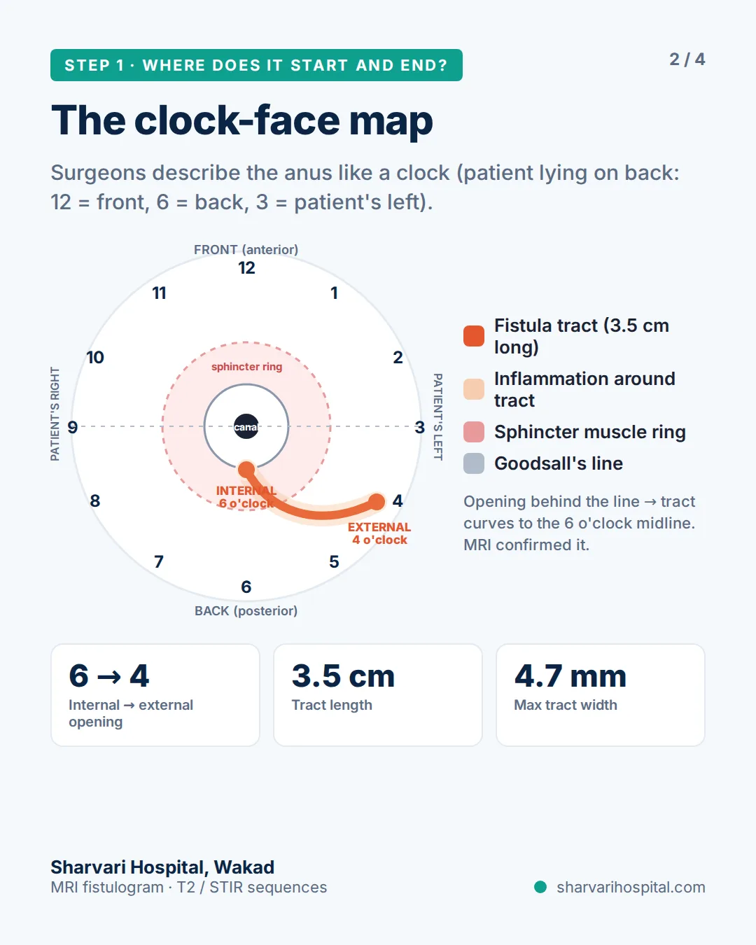
Almost every fistula patient at Sharvari Hospital is imaged before surgery — an MRI fistulogram or a trans-rectal ultrasound. Because the external opening sat behind Goodsall’s line, where the tract usually curves to the posterior midline, an MRI fistulogram was requested to confirm the route and the depth before any decision about the operation.
MRI fistulogram findings
| Finding | MRI report (identity removed) |
|---|---|
| Internal opening | 6 o’clock (posterior midline of the anal canal) |
| External opening | 4 o’clock (left, behind the anus) |
| Tract | Single tract, 3.5 cm long, 4.7 mm at its widest, curving through the left ischioanal fossa |
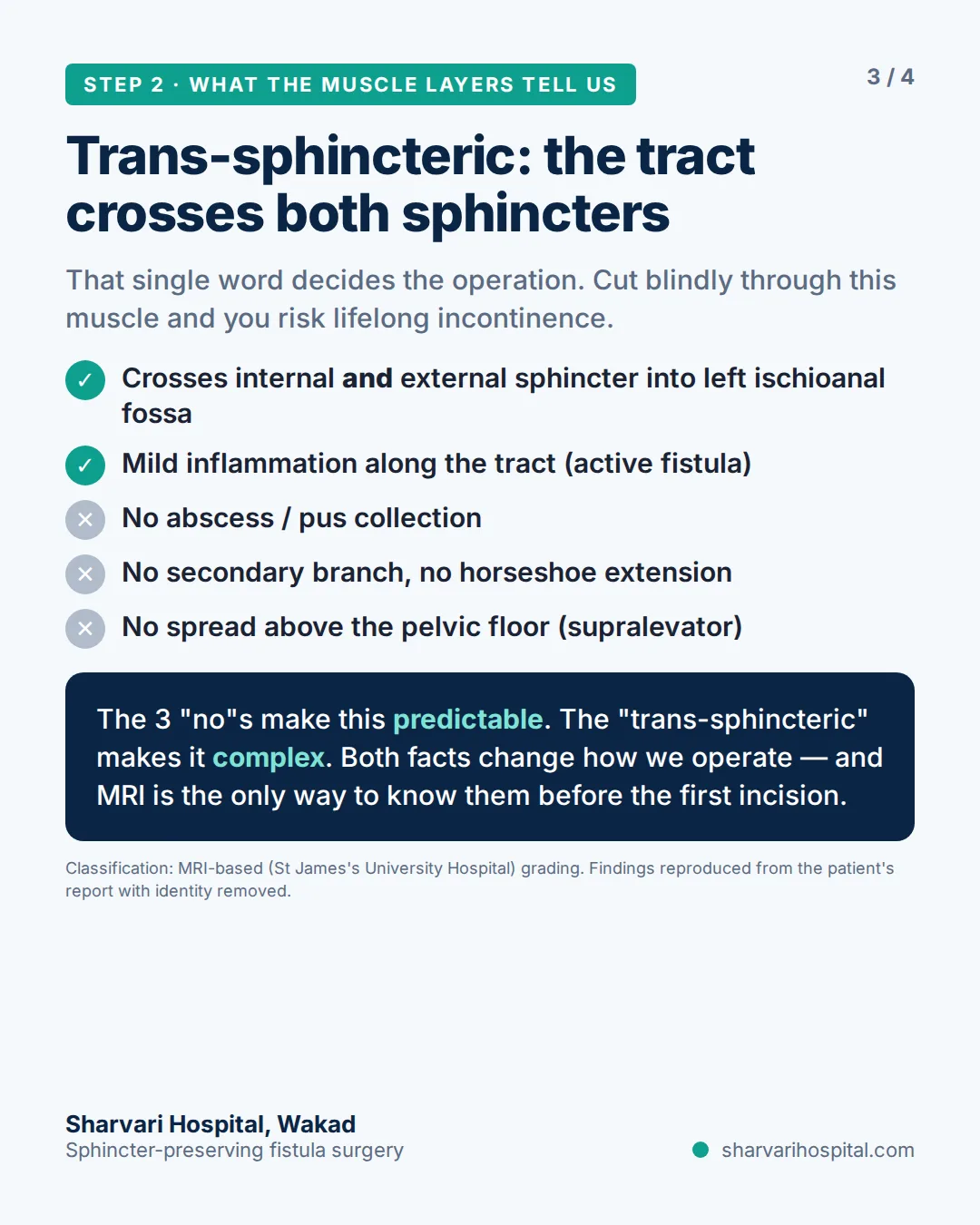
| Relation to the sphincters | Trans-sphincteric — crosses both the internal and the external sphincter |
| Inflammation | Mild inflammation along the tract (active fistula) |
| Abscess | None |
| Secondary tract / horseshoe | None |
| Supralevator extension | None |
Three “no” findings — no abscess, no secondary tract, no supralevator extension — made this a predictable single tract. One word — trans-sphincteric — made it complex. Both facts changed how the operation was done. How to read each line of a report is explained on the MRI fistula mapping page.
What the map changed in the plan
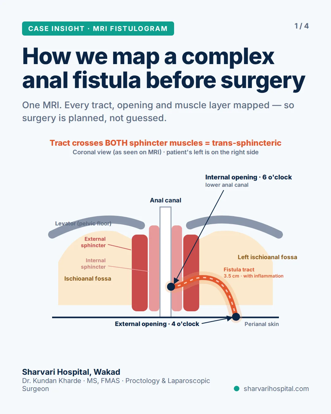
A single, short posterior tract in a young man with no previous surgery would usually be treated by fistulotomy — laying the whole tract open so it heals flat. That is the operation with the lowest chance of the fistula returning.
The MRI showed why that was not safe here. The tract crossed both the internal and the external sphincter on its way to the skin. Opening the entire length would have divided the muscle that controls stool.
So the plan changed from a full lay-open to a hybrid: lay open the part of the tract that is safe to open, and seal the part running through the external sphincter with laser (FiLaC) so that muscle is never cut. The decision was made from the scan, before the first incision, and then checked in theatre.
In the operating theatre: the probe follows the map
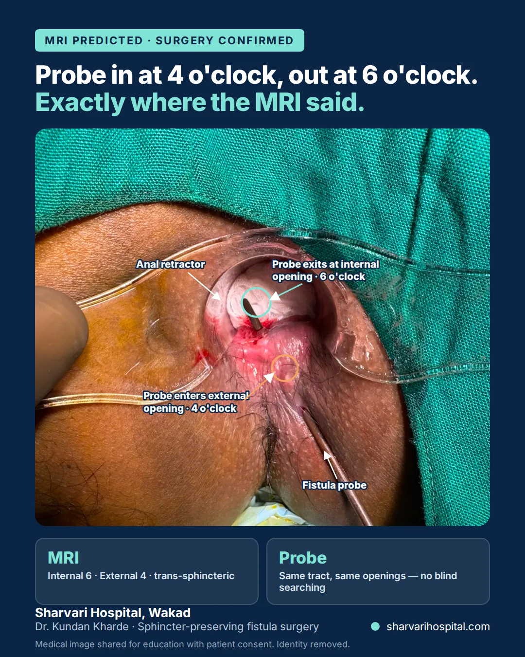
Under anaesthesia, a fistula probe was passed into the external opening at 4 o’clock. It travelled along the tract and came out at the internal opening at 6 o’clock — the same tract, the same two openings, exactly as the MRI had drawn it. There was no blind searching for the internal opening, no false passage and no need to explore for a branch that the scan had already ruled out. The photograph shows the probe in place with the anal retractor open, the tip visible at 6 o’clock inside the canal.
The operation: fistulotomy for the proximal tract, laser for the part inside the external sphincter
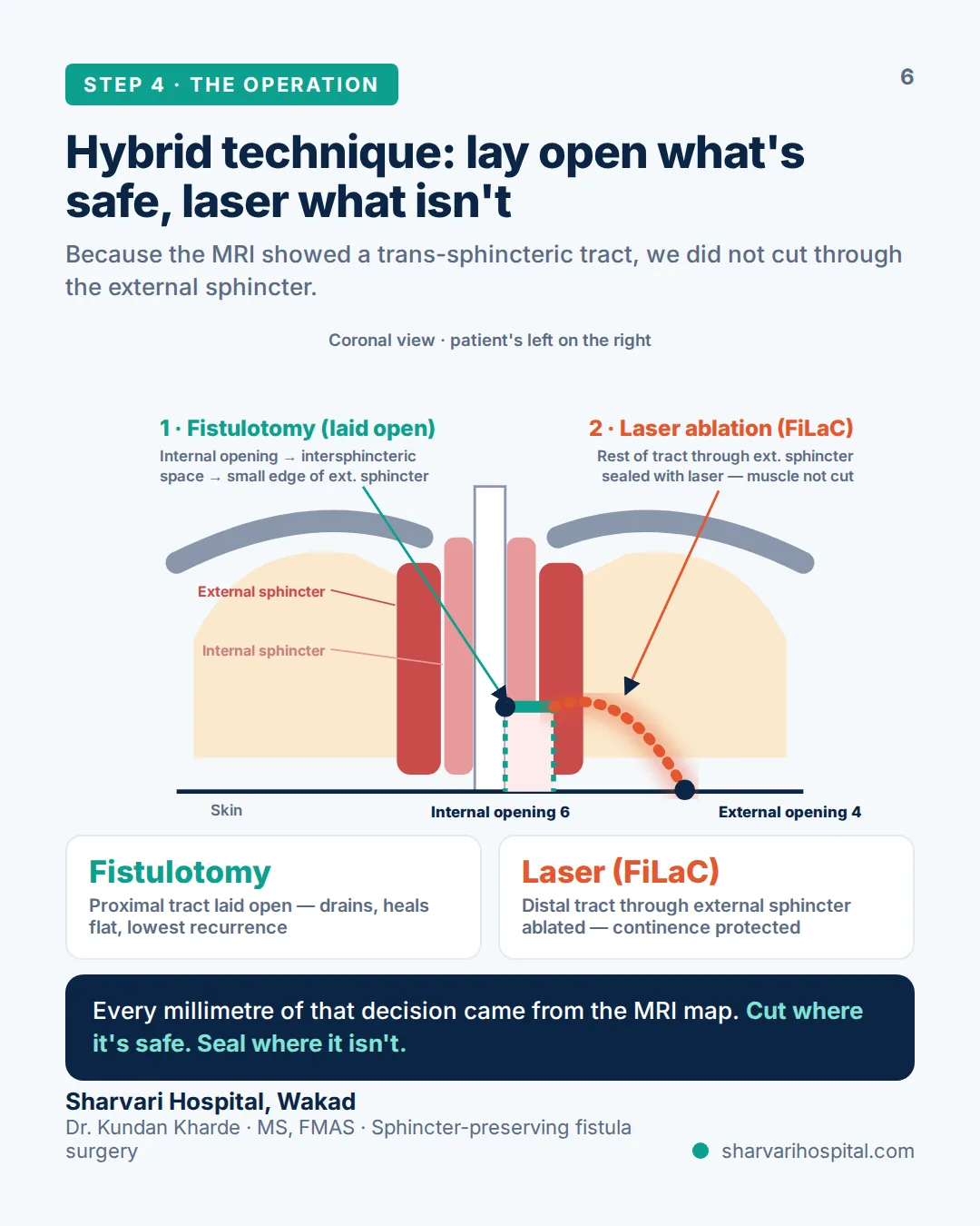
Step 1 — fistulotomy of the proximal tract. The tract was laid open from the internal opening at 6 o’clock through the intersphincteric space, taking only a small edge of the external sphincter. This part drains freely, heals flat from the base and is the segment where recurrence usually starts if it is left closed.
Step 2 — FiLaC for the remainder. The rest of the tract, running through the external sphincter to the 4 o’clock external opening, was not cut. A radial laser fibre was passed along it and the tract lining was ablated and sealed from inside, so the muscle around it stays intact.
Why the hybrid. Continence protection is the whole rationale. Cutting through the external sphincter to lay open a trans-sphincteric tract risks lifelong control problems; sealing the muscle-bearing part with laser avoids that while still dealing with the internal opening properly. Every millimetre of that decision came from the MRI map.
Recovery
Post-operative images and follow-up will be added at the patient’s review.
What this case shows
A fistula that looked simple from outside was trans-sphincteric on MRI, and the scan changed the operation before the first incision. The probe in theatre confirmed the map exactly, so nothing was searched for blindly. The part of the tract inside the external sphincter was sealed with laser rather than cut, which is how continence was protected.
Consent and privacy
This case is published for patient education with the patient’s written consent. No name, patient identifier, scan date, report or radiologist details are shown; the MRI findings are summarised in plain words and diagrams rather than reproduced from the report. The intra-operative photograph has been cropped so that no identifying feature is visible. Every case study on this site follows the same rule: the anatomy and the decision are shared, the person is not.
Already have an MRI? Get Dr. Kharde’s opinion on it
WhatsApp the written report and photos of the key images to +91 951 951 1928. Dr. Kundan Kharde reads it before you travel and tells you what the findings mean for the operation.
Fistula that has come back after surgery is planned on its own page: complex & recurrent fistula treatment. First-time patients start at fistula treatment in Pune. How the map is read: MRI fistula mapping.
Your surgeon: Dr. Kundan Kharde, MS, FMAS — Founder & Director, Lead Proctology Surgeon, Sharvari Hospital. 19+ years, 6,000+ surgeries.
SR. NO. 19(P), behind Gulmohor Park, Vishal Nagar, Pimple Nilakh, Pune 411027 · Open 24 hours · +91 951 951 1928