Case studies · Recurrent fistula · MRI-mapped · De-identified
Case study: recurrent complex fistula — the MRI found a 6 cm abscess that could not be seen from outside
A fistula operated elsewhere about eight years ago came back as eight days of pain and one small opening near the anus. From outside there was almost nothing to see. The MRI fistulogram showed a 6 cm horseshoe abscess and a tract crossing the external sphincter — and that map decided a sphincter-preserving fistulectomy rather than a full lay-open or a surface clean-up.
Presentation
A man in his early thirties came to Sharvari Hospital, Pimple Nilakh, with eight days of pain around the anus. He had been operated for a fistula at another hospital about eight years earlier and had been well since. There was no significant discharge — no staining, no pus that he had noticed — only the pain.
On examination there was a single small external opening near the anus, in the midline behind it, with scar from the previous operation around it. There was no obvious swelling, no redness spreading across the buttock and nothing on the surface that pointed to a large collection.
What the examination could tell: that the old fistula had reopened, and roughly where the tract met the skin. What it could not tell: where the tract started inside the anal canal, how much sphincter muscle it crossed, and whether the pain was coming from something larger hidden in the deeper tissue. In a fistula that has already been operated on, scar makes all three questions harder to answer with a finger. Almost every fistula patient at Sharvari Hospital is imaged before surgery — an MRI fistulogram or a trans-rectal ultrasound. Here an MRI fistulogram was requested before any decision about the operation.
What the MRI fistulogram showed
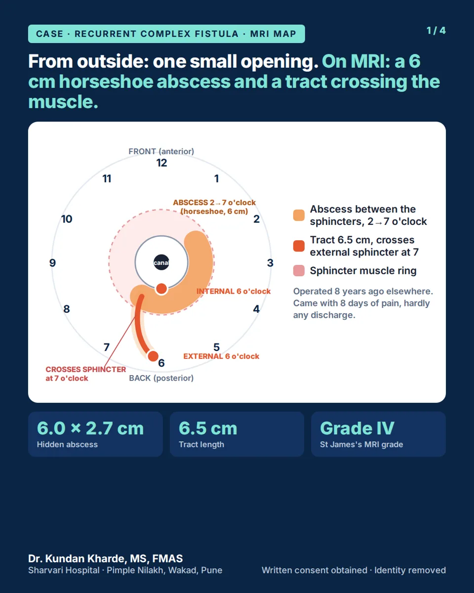
The MRI fistulogram (plain, without contrast) was done before surgery. The findings, in plain words, are set out in the table below. The number that mattered most was 6 cm: a collection that size, spread in a horseshoe behind the rectum, was producing eight days of pain and yet almost no discharge, because it was draining only through a long, narrow tract to one small skin opening.
What grade IV means. In the St James’s University Hospital MRI classification, grade IV is a trans-sphincteric tract with an abscess or a secondary tract in the space outside the sphincter — the second-highest grade, one below tracts that climb above the pelvic floor.
| Finding | MRI fistulogram (identity removed) |
|---|---|
| Abscess | Large multilobulated collection, about 6 × 2.7 × 2.1 cm, in the posterior perirectal and intersphincteric space — behind the rectum, between the two sphincter layers |
| Horseshoe spread | The collection wrapped around from about 2 o’clock to 7 o’clock |
| Internal opening | Probable internal opening at 6 o’clock (posterior midline of the anal canal) |
| External opening | Right gluteal cleft at 6 o’clock — the single small opening seen on examination |
| Tract | About 6.5 cm long, from the external opening up to the abscess |
| Relation to the sphincters | Crosses the right external sphincter at about 7 o’clock — trans-sphincteric |
| Secondary tracts | None |
| Supralevator extension | None — nothing above the pelvic floor |
| MRI grade | St James’s University Hospital grade IV |
Two “no” findings — no secondary tract, no supralevator extension — kept this within reach of a single operation. Two “yes” findings — a 6 cm horseshoe abscess and a tract crossing the external sphincter — decided what that operation had to be. How to read each line of a report is explained on the MRI fistula mapping page.
Why this changes the operation
With this map in hand, two operations that would have looked reasonable from outside were ruled out.
A simple lay-open of everything — following the tract from the skin to the internal opening and opening it along its whole length — would have cut through the right external sphincter where the tract crosses it at about 7 o’clock. That is the muscle that controls stool, and in a patient who has already had one fistula operation there is less of it to spare.
A blind “clean-up” of the outside opening — excising the skin opening and scraping whatever tract can be felt from outside — would have left the 6 cm abscess and the internal opening at 6 o’clock behind. The tract would have refilled from the inside, and the fistula would have come back for a second time. An internal opening that is never dealt with, and an intersphincteric collection that is never drained, are the two most common reasons a fistula recurs.
So the plan was built in two parts, from the scan and before the first incision: deal with the source and the abscess from inside, and remove the part of the tract crossing the muscle without dividing the muscle.
In the operating theatre
The operation was a fistulectomy — the tract was removed rather than only cut open. The two photographs below are explicit surgical images and are blurred until you choose to view them.

Core-out of the part crossing the muscle. Under anaesthesia the internal opening at 6 o’clock, the intersphincteric space and the abscess cavity behind the rectum were laid open and drained, and the horseshoe extension was cleared from within. The part of the tract crossing the right external sphincter at about 7 o’clock was then cored out — removed as a cylinder of tract tissue — leaving the muscle around it uncut.

Whole tract removed to the skin opening. The cored-out tract was followed outward to the skin and the whole length of it was removed rather than only opened. The external opening in the gluteal cleft, with its rim of scar from the previous surgery, was excised with it, so no lined tract was left behind to reopen.
Why “core out” instead of “cut”
Cutting a tract open (fistulotomy) divides everything between the tract and the skin — including any sphincter muscle that lies in the way. Coring out removes the tract itself as a cylinder from inside the muscle and leaves the muscle fibres around it intact. In this operation the external sphincter was not divided.
Fistulectomy means the tract was removed, not just opened. The distinction matters in a recurrent fistula: a tract that has been there for years has lined itself with skin-like tissue that will not stick together and close, and removing it takes that lining out of the wound. The sphincter-preserving part of the plan — coring rather than cutting where the tract crosses the muscle — is what protects bowel control. The general principles are on the complex & recurrent fistula treatment page.
Pain relief after surgery

At the end of the operation a long-acting local anaesthetic (bupivacaine) was infiltrated around the wound as a block, for pain relief in the first hours after surgery. It was mixed with a small amount of methylene blue dye so the surgeon can see exactly where the block has been given — the blue staining in this photograph is that dye, not staining of the tract.
He was discharged the next day, and the wound was healing at follow-up. That is this patient’s course; healing after a fistulectomy differs from person to person and no timeline is promised.
Why recurrent fistulas need mapping first
A fistula that has already been operated on behaves differently from a first fistula. Scar hides the internal opening, the tract may have taken a new route, and — as here — a collection can build up in the deep tissue with almost nothing to show on the surface. Examination alone answered none of the questions that decided this operation.
An MRI fistulogram answers them before the first incision: where the tract starts, how much sphincter it crosses, and whether there is an abscess, a second tract or a horseshoe extension to deal with. How the map is read is on the MRI fistula mapping page; how a fistula that has come back is re-assessed and planned is on complex & recurrent fistula treatment. A recurrent fistula is not re-operated at Sharvari Hospital without that map.
What this case shows
Eight days of pain and one small opening hid a 6 cm horseshoe abscess and a tract crossing the external sphincter. The MRI found what the examination could not, and it ruled out two operations that would have looked reasonable from outside: a full lay-open that would have cut the muscle, and a surface clean-up that would have left the abscess and the internal opening behind. The abscess and the internal opening were dealt with from inside; the part of the tract crossing the muscle was cored out rather than cut; the whole tract was removed to the skin. He went home the next day. This is one patient’s case, not a statistic and not a promise.
Consent and privacy
This case is published for patient education with the patient’s written consent for both the images and the MRI findings. No name, patient identifier, scan date, report or radiology-centre details are shown; the MRI findings are summarised in a table and a diagram, and the report itself is not reproduced. The intra-operative photographs have been cropped so that no identifying feature is visible, and they are shown only after you choose to view them. Every case study on this site follows the same rule: the anatomy and the decision are shared, the person is not.
Already have an MRI? Send the report for Dr. Kharde’s opinion
If your fistula has come back after surgery, WhatsApp the written MRI report, photos of the key images and your previous operation notes to +91 951 951 1928. Dr. Kundan Kharde reads them before you travel and tells you what the findings mean for the operation.
Fistula that has come back after surgery is planned on its own page: complex & recurrent fistula treatment. How the map is read: MRI fistula mapping. Dr. Kharde’s own page on recurrent fistula: complex & recurrent fistula — drkundankharde.com.
Your surgeon: Dr. Kundan Kharde, MS, FMAS — Founder & Director, Lead Proctology Surgeon, Sharvari Hospital. 19+ years, 6,000+ surgeries.
Frequently asked questions
Can a fistula come back years after surgery?
Yes. A fistula can recur many years after an operation, as it did in this case about eight years later. The usual reasons are an internal opening that was not dealt with, a branch or intersphincteric collection that was left undrained, or a tract that closed at the skin but not in the depth. A recurrence is a reason to map the tract on MRI before any second operation, not a reason to repeat the first one.
Why did the MRI find an abscess when there was hardly any discharge?
Because the abscess was deep — behind the rectum and between the sphincter layers — and its only route to the skin was a long, narrow 6.5 cm tract ending in one small opening. Very little pus reached the surface, so there was pain but almost no discharge. Discharge reflects how freely a collection drains, not how large it is, which is why a painful recurrence with little discharge still needs imaging.
What does "St James's grade IV" mean on an MRI report?
The St James’s University Hospital classification grades an anal fistula from 1 to 5 on MRI. Grade IV means a trans-sphincteric tract — one that crosses both the internal and the external sphincter — together with an abscess or a secondary tract in the space outside the sphincter. Grade V is reserved for tracts that extend above the pelvic floor. The grade is a radiology shorthand; the operation is planned from the actual findings and confirmed under anaesthesia.
Will fistula surgery affect bowel control?
The risk to bowel control comes from cutting the external sphincter, and it rises with each previous operation. That is why, in this case, the part of the tract crossing the external sphincter was cored out — removed as a cylinder from within the muscle — rather than cut through, and the muscle was left intact. No fistula operation has zero risk, so the technique is chosen from the MRI and the amount of muscle involved, and it is discussed with you before consent.
Do I need an MRI before redo fistula surgery?
Yes. Almost every fistula patient at Sharvari Hospital is imaged before surgery, and for a recurrent fistula the MRI fistulogram is the starting point rather than an optional test. Scar from the previous operation hides the internal opening and the true route of the tract from the examining finger, and a deep collection can be present with little on the surface, as in this case. The scan shows where the tract starts, how much sphincter it crosses and whether an abscess or a second tract has to be dealt with, and the operation is planned from that map.
SR. NO. 19(P), behind Gulmohor Park, Vishal Nagar, Pimple Nilakh, Pune 411027 · Open 24 hours · +91 951 951 1928